A quick history of fetal monitoring: Auscultation ~ verbs, nouns, and names of physician-inventors
 The first person to report hearing the sound of an unborn baby’s heartbeat was a 17th century physician by the name of Dr. Marsac. A contemporary of his, a Dr. Killian, was the first to ponder the possibility that the rate and rhythm of the fetal heart might be an indicator of how well the fetus was (or wasn’t) tolerating the process of childbirth.
However, this possibility went unnoticed for two more centuries, until 1818, when doctors Mayor and Kergaradec described a method of auscultating fetal heart sounds by placing one’s ear on or very near the maternal abdomen. Dr. Kergaradec also suggested that fetal heart sounds could be used to determine fetal viability and proof of life in the months before ‘quickening’, that is, before the mother could feel the baby kick.
In 1833, an English physician, Dr. Evory Kennedy, published guidelines for fetal distress and also recommend auscultation of the fetal heart rate to monitor the well-being of the fetus during labor and birth.
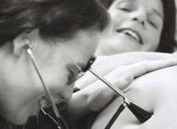
A simple tool for counting the fetal heart rate
However, the big leap forward came in 1893, when Dr. Von Winkel established criteria for fetal distress. After listening to the heart rate of many babies during many labor, he realized that the FHR of an unborn baby in trouble would often (but not always) become abnormal, and beat much faster or much slower, or become extremely erratic. Some of these babies died before the birth, other were born profoundly depressed and often died later.
With this understanding, Dr Von Winkel identified the signs of fetal distress,which included abnormal fetal heart rates (FHR):
- tachycardia (a over 160);
- bradycardia (under 100),
- irregular heart rate, passage of meconium,
- abnormal fetal movement (i.e. agonal spasms of a dying baby).
By the turn of the 20th century, maternal fever was recognized as a cause of fetal tachycardia; head compression and cord compression were also known causes of bradycardia, and hyperstimulated uterine activity associated with an abnormal FHR patterns and asphyxia.
When these abnormal heart rates were discovered very late in the labor, doctors could sometimes perform a forceps delivery to rescue the baby. By the late 19th century, the development of general anesthesia, germ theory of infectious disease (resulting the principles of asepsis and sterile surgicaltechniques) made Cesarean surgery safe enough to use during labor to rescue babies with extremely abnormal heart rates.
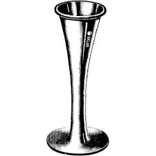
 By the early 20th century — 50 years before fetal heart monitoring was electronically automated by a computerized machine — doctors, midwives and nurses were using a special stethoscope called a Pinard Horn to regularly listen to the heartbeat of the fetus during labor. Pinard fetal stethoscope was developed in the 1880s and in wide use in the 1950s.
Helpful as this information was, there were still a few babies with apparently “normal” FHR that were born with unexpected and unexplained problems that prevented the baby from breathing on its own that resulted in death or severe handicaps.
At this early stage of development of fetal monitoring, auscultation was only used to count the heart rate for one minute (b.p.m, charted as a baseline for that period of time). However, birth attendants and nurses paid no attention to the rhythm of the fetal heart or other elements now known to be equally, if not even more important — variability, the presence of reassuring accelerations and absence of pathological decelerations, as well as the depth, timing, and duration of any decelerations that are detected.
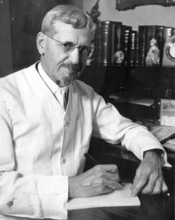
 However, in 1922 Dr. Joseph De Lee, obstetrician, founder of the Chicago Lying-in Hospital and Chicago Maternity Center, invented, in conjunction with another obstetrician (Dr. David Hills) a hands-free fetal stethoscope for listening to fetal heart tones. This device became known as the DeLee-Hills Fetoscope.
Then Dr. DeLee developed the process and protocols for the intermittent auscultation (IA) of the fetal heart rate during labor, which included Dr. Von Winckel’s 1893 description of fetal distress listed above. Using his new fetoscope, the FHR was to be auscultated every 30 minutes during first stage of labor, every three or five minutes during second stage, and continuously if signs of fetal distress were seen as an indication for forceps delivery.
In 1924, 4th edition of Dr. DeLee’s obstetrical textbook Principles and Practice of Obstetrics includes a wonderful graphic that displays a fetal heart rate auscultated and charted in 5-second increments during a minute-long contraction.
 It shows a normal baseline rate of 132 bpm at the beginning of the contraction (ranging from 120 to 144 bpm when computed in 5-second samplings) during the first 20 seconds. As pressure on the fetal head builds up, there is a head compression decel, and we can see the FHR go down from 144 to 108 bpm over a span of 20 second durning the middle of the contraction. As the contraction wanes and the uterus relaxes during the last 20 seconds, the unborn baby’s heart rate speeds back up to its pre-contracton normal baseline of 132.
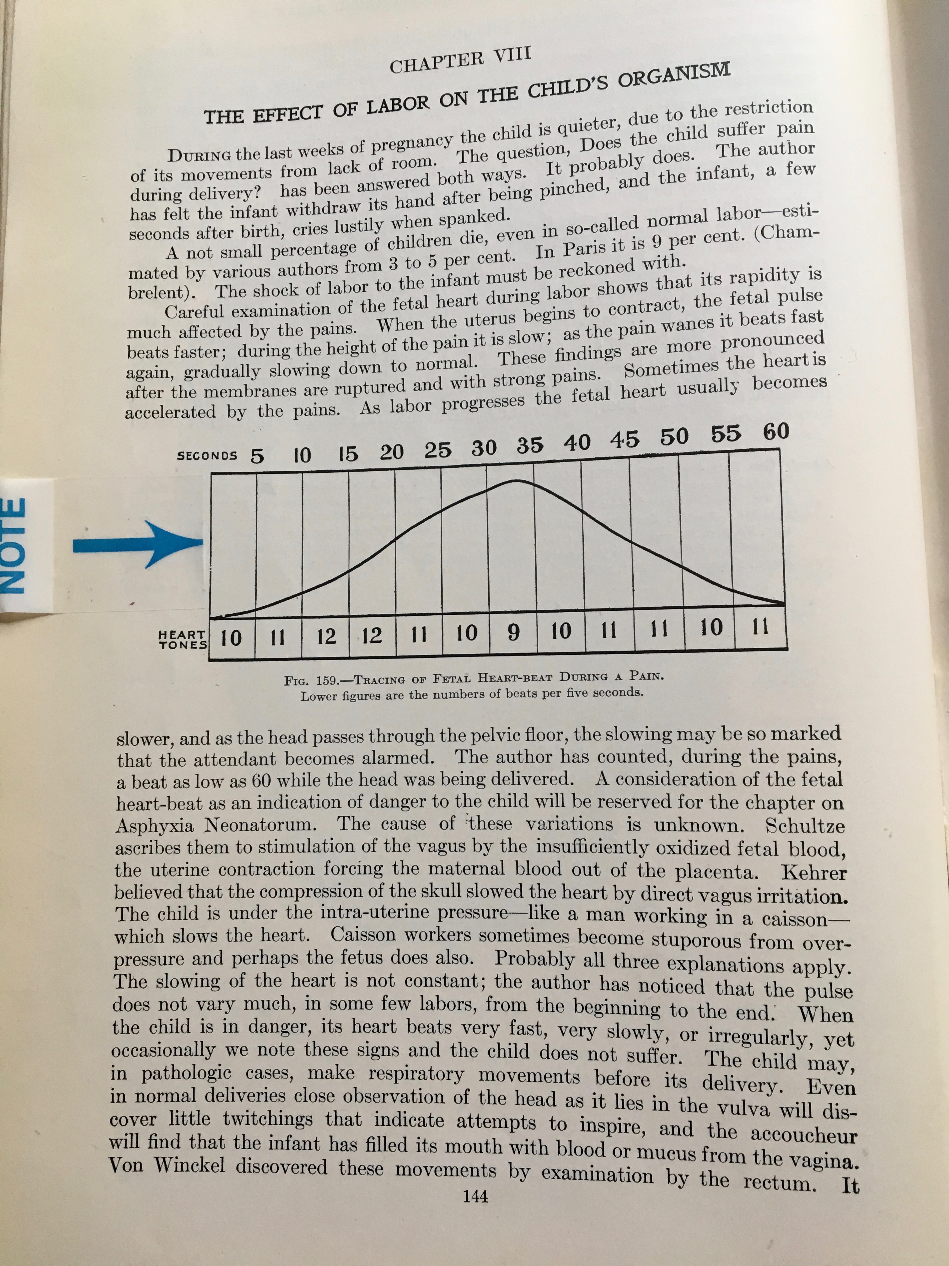
 If you read Dr. DeLee’s comments below the picture of the graph, he refers to a pattern of decelerations during the pushing stage that we now know to be head compression decels — the result of triggering the mammalian diving reflex that has been extensively studied in whales.
Dr. DeLee somewhat anticipated this explanation by referring to the observed effect of an abnormally slow heart rate of men working deep under water in a “caisson” (a big metal contraption used during the underwater building of bridges and similar construction projects).
DeLee’s work is the theoretical precursor to both IA and EFM, as it is the first instance of dealing with variations in the FHR during the period of auscultation by using 5-second samplings. Prior to this, the only information on fetal heart activity that was gathered was the sum total of beats over 60 seconds period, which was stated as beats per minute (bpm) and recorded as a baseline rate.
Dr. DeLee recognized a second-level source of valuable information that could be gathered by paying attention to the FHR changes that occured (or failed to occur) within the one- or two-minute period that the FHR were routinely monitored by the L&D nurse, doctor or midwife.
The theoretical basis developed by Dr. DeLee and its 5-second sampling protocol, which yielded much a more useful picture of fetal wellbeing. This critical aspect of IA was widely not acknowledged or integrated into standard obstetrical practice until the development of electronic fetal monitoring equipment in the late 1950s and 60s.
If you are interested in learning how auscultations works, this link will take you to an educational post
Note: The historical background material on non-electronic fetal monitoring is repeated. To skip that part, keep scrolling down until you see this photo and the heading:”How IA Works“.
Next post in MAYDAY series: The invention of electronic automated fetal monitors and its displacement of auscultation/hands-on listening as a part of the laboring woman’s one-on-one care by a professionally-trained labor and birth attendant